Rat Dissection
Purpose
To investigate the external and internal structures of a chordate, specifically a rat.
Materials
Rat specimen
Dissecting tray
Dissecting kit
Dissecting tray
Dissecting kit
Method - Part 1: External anatomy
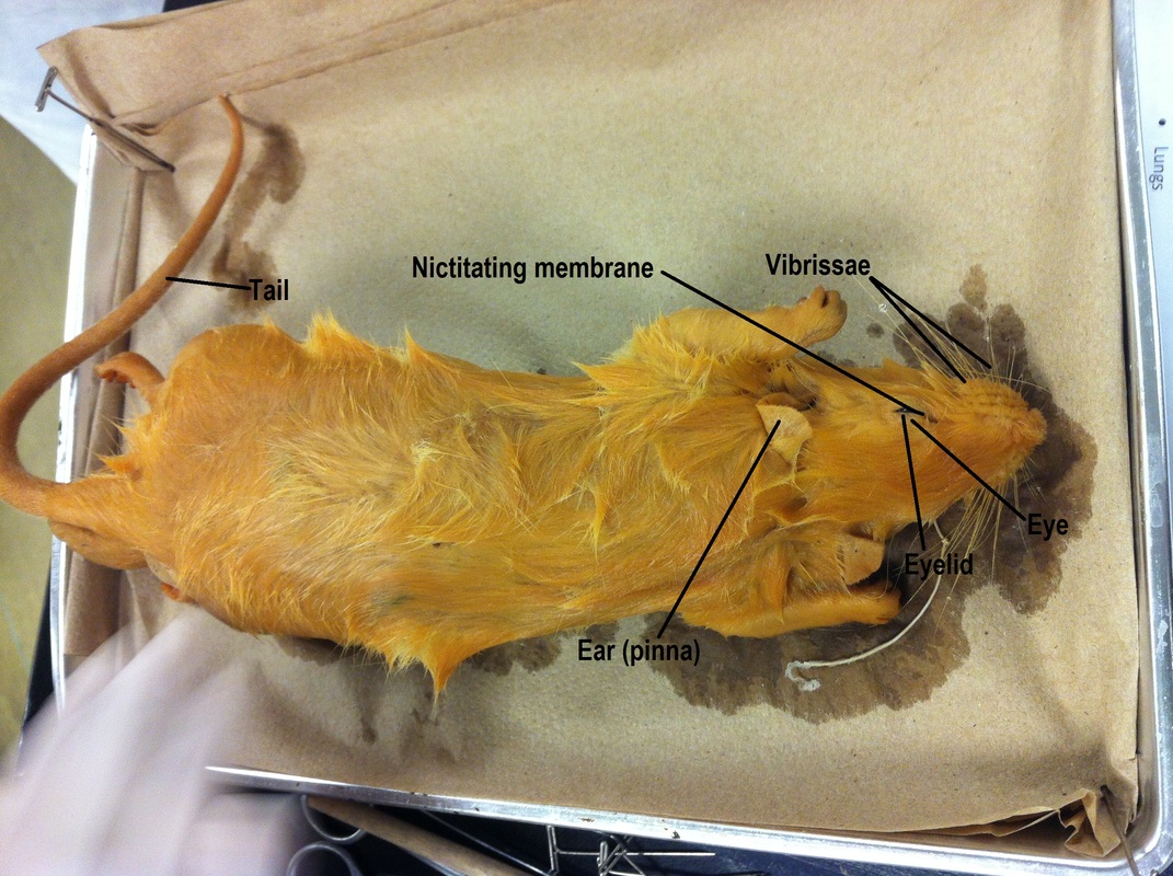
1. Name your rat! Our rats name is Juan (spanish, J is prononced as a "wh" sound)
2. Obtain your rat and observe the general characteristics. The rats body is divided into six anatomical regions; the cranial region, head cervical region, neck pectoral region, area where front legs attach thoracic region, chest area abdomen, belly pelvic region, and the area where the back legs attach.
3. Note the hairy coat covering the rat and the sensory hairs (whiskers) located on the rats face, called vibrissae.
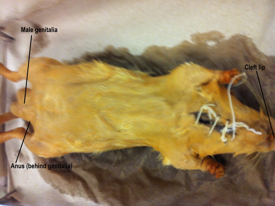
4. The mouth has a large cleft in the upper lip which exposes the large from incisors. Rats are gnawing mammals, and these incisors continue to grow as long as the rat lives.
5. Note the eyes with large pupils and nictitating membrane found at the inside corner of the eye. This membrane can be drawn across the eye for protection. The eyelids are similar to those found in humans.
6. Locate the teats on the ventral surface. Check a rat of another sex and determine whether both sexes have teats.
7. Examine the tail, of which on rats have no hair. Though some rodents, like gerbils, do.
8. The ears are composed of the external part, called the pinna, and the auditory meatus, the ear canal
9. Locate the anus, which is ventral to the base of the tail.
10. Determine whether your rat is male or female by looking near the base of the tail for male or female genital organs.
Part 2: Skinning the rat
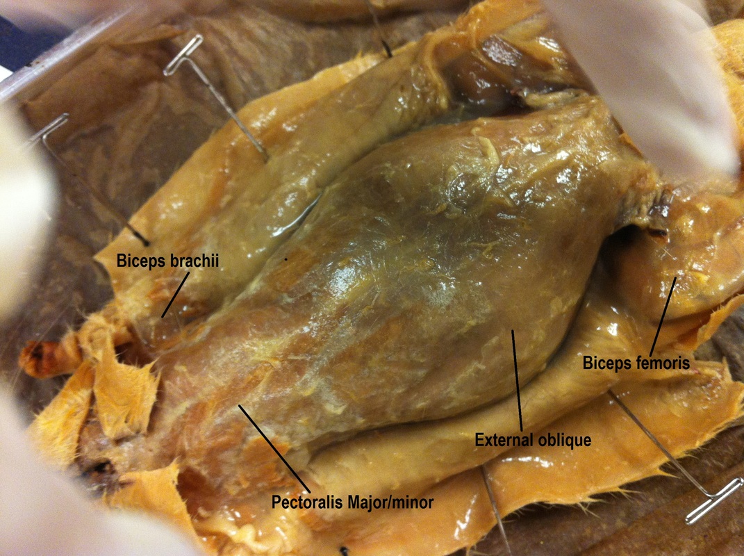
1. You will carefully remove the skin of the rat to expose the muscle below. This task is best accomplished by making a small incision with a scalpel and using a probe to seperate the connective tissues connecting the skin to the first layer of muscles. Do not cut the muscles!
2. Start the incision where the latex was injected and continue towards the tail, avoiding the genital area. Gently peel the skin away from the muscles, using scissors and a probe to tease away muscles that stick to the skin.
3. Identify the following muscles: Biceps brachii - located on the anterior surface of the arm; flexes lower arm. Biceps femoris - Located on side of thigh, in two bundles; flexes lower leg. External Oblique - Located on side of abdomen; flexes body wall. Pectoralis major - Located in chest; draws arm foreward.
Part 3: Skeletal system
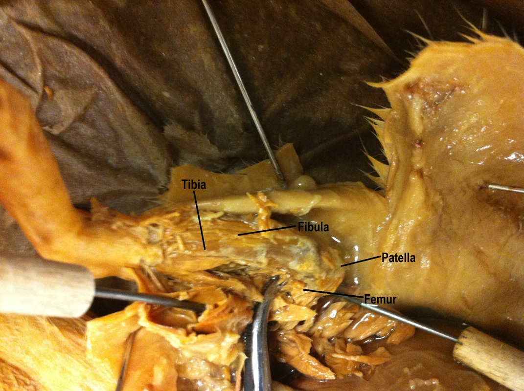
1. Cut away the muscle to expose the bones of the leg. Carefully tease away the bicep femoris and gastrocnemius on one leg to expose the 3 leg bones: Tibia, Fibula, and femur, with the small patella (Knee cap).
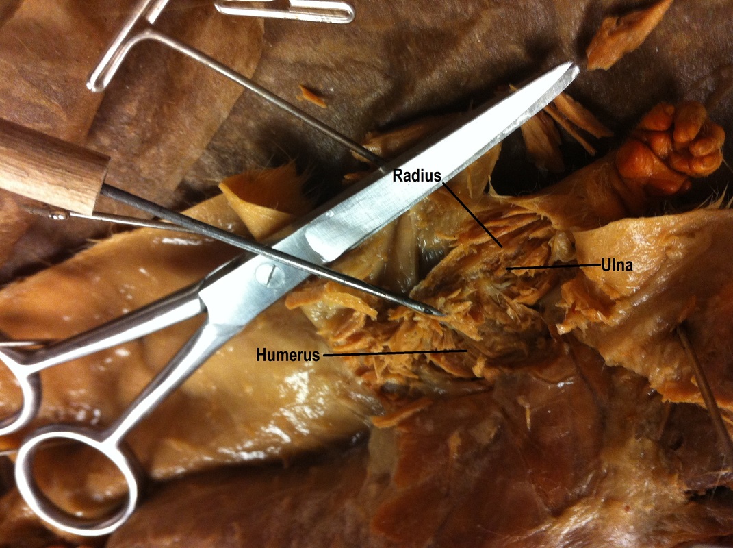
2. You can also see the ligaments around the knee that attach the bones of the lower leg to the femur and the achilles tendon which attaches the gastrocnemius to the ancle. Remove the muscles from one arm to reveal the ulna, radius, and humerus. Note the size of the radius.
Part 4: Digestive system
1. Use scissors to cut through the abdominal wall of the rat following the insicion marks made when skinning. Be careful not to cut too deeply, keeping the tip of your skalpel pointed upwards. Do not damage the underlying strucures.
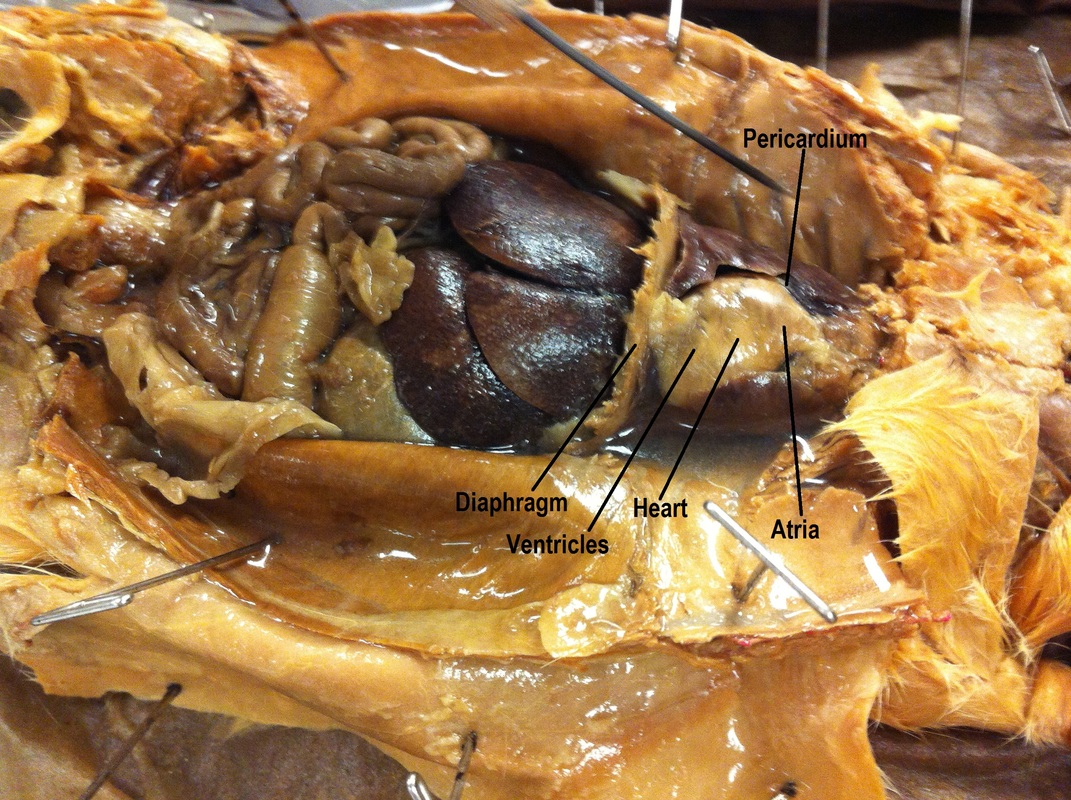
2. Locate the diaphragm, a thin layer of muscle seperating the thoracic cavity from the abdominal cavity. The diaphragm is a helful directional marker for the rest of the dissection.
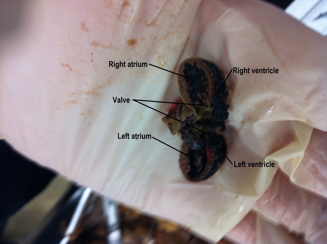
3. Do not remove or cut the heart! The heart is centrally located in the thoracic cavity. The two dark-coloured chambers at the top are the atria, and the bottom chambers are the ventricles. The heart is covered by a thin membrane called the pericardium.
Part 5: Abdominal organs
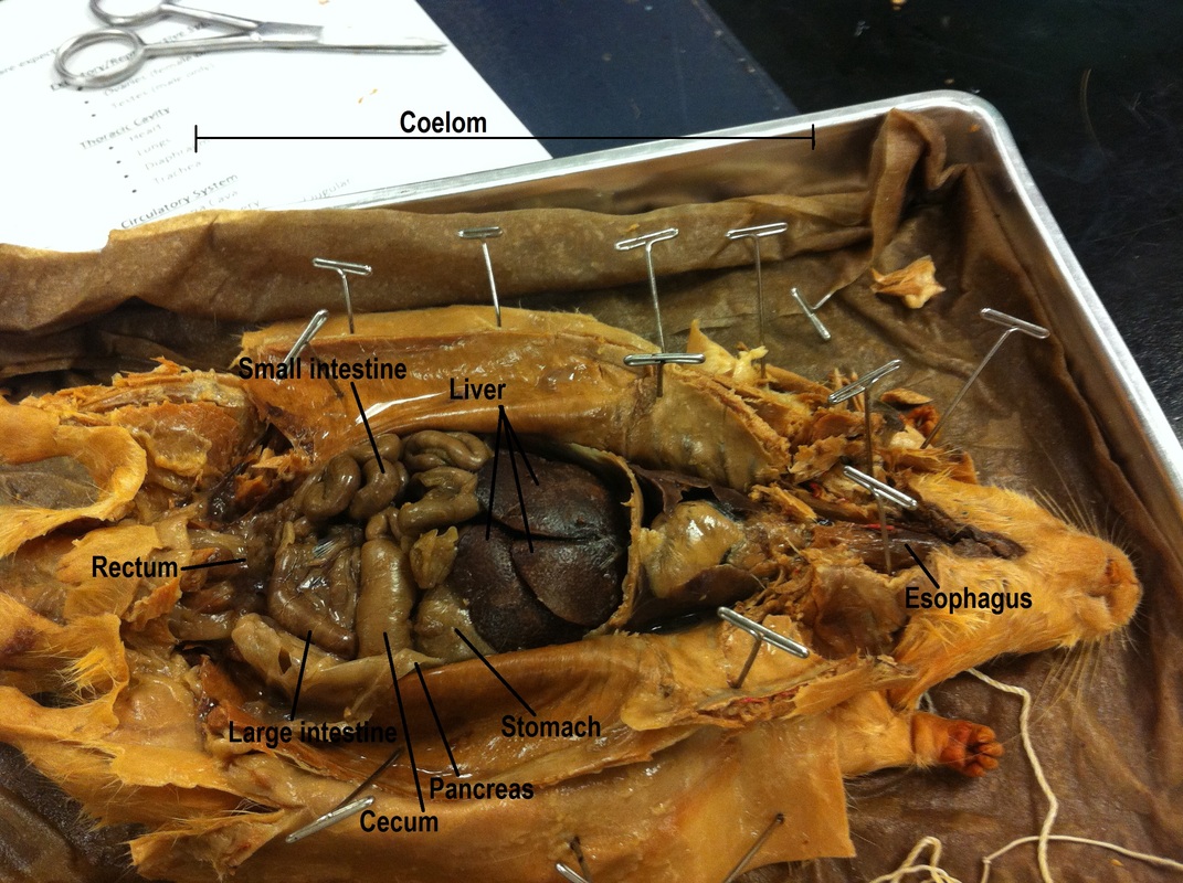
1. The coelum is the body cavity within which the viscera (internal organs) are located. The cavity is covered by a membrane called the peritoneum.
2. Locate the liver, a large dark coloured organ suspended above the diaphragm. The liver has many functions, one of which is to produce bile which aids in digesting fat. The liver also store glycogen and transforms waste into less harmful substsances. Rats dont have a gall bladder which is used for storing bile in other animals.
3. The esophagus runs through the diaphragm and moves food from the mouth to the stomach. It is distinguished from the trahcia by its lack of cartilage.
4. Locate the stomach on the right side just under the liver. The functions of the stomach includes food storage, physical breakdown of food, and digestion of protein. The opening between the esophagus and the stomach is called the cardiac sphincter. The outer margin of the stomach is called the greater curvature, the inner margin is called the lesser curvature.
5. The spleen is about the same colour as the liver and is attatched to the greater curvature of the stomach. Its shaped like a banana and is associated with the circulatory system and functions as the destruction of blood cells and blood storage. A person can live without a spleen, but they're more likely to get sick as it helps the immune system.
6. The pancreas is not a clearly identifiable organ but a thin membrane overlaying the stomach and spleen. The pancreas produces digestive enzymes sent to the intestine via small ducts. It also secretes insulin, which is important in the regulation of glucose metabolism.
7. The small intestine is a slender coiled tube that recieves partially digested food from the stomach. The term small refers to its diameter, not its length. It consists of three sections; duodenum, ileum, and jejunum. The small intestine leads to the cecum.
8. The cecum is a pouch connecting the large and small intestines. Food is temporarily stored in the cecum while bacteria digest cellulose found in plants. Most herbivores have a large cecum, while humans and other carnivors and omnivores have a smaller cecum called the appendix.
9. Use your scissors to cut the mesentery of the small intestine, but do not remove the small intestine from the rectum and stomach. You can stretch it out and untangle it so that you can see the relative lengths of the large and small intestine.
10. Locate the large intestine, which is the large greenish tube that extends from the small intestine to the anus. It is also known as the colon. This is where the final stages of digestion and water absorption occurs.
11. Locate the rectum - the short, terminal section of the colon between the end of the large intestine and the anus. It temporarily stores feces before they are expelled from the body.
Part 6: Excretory systems
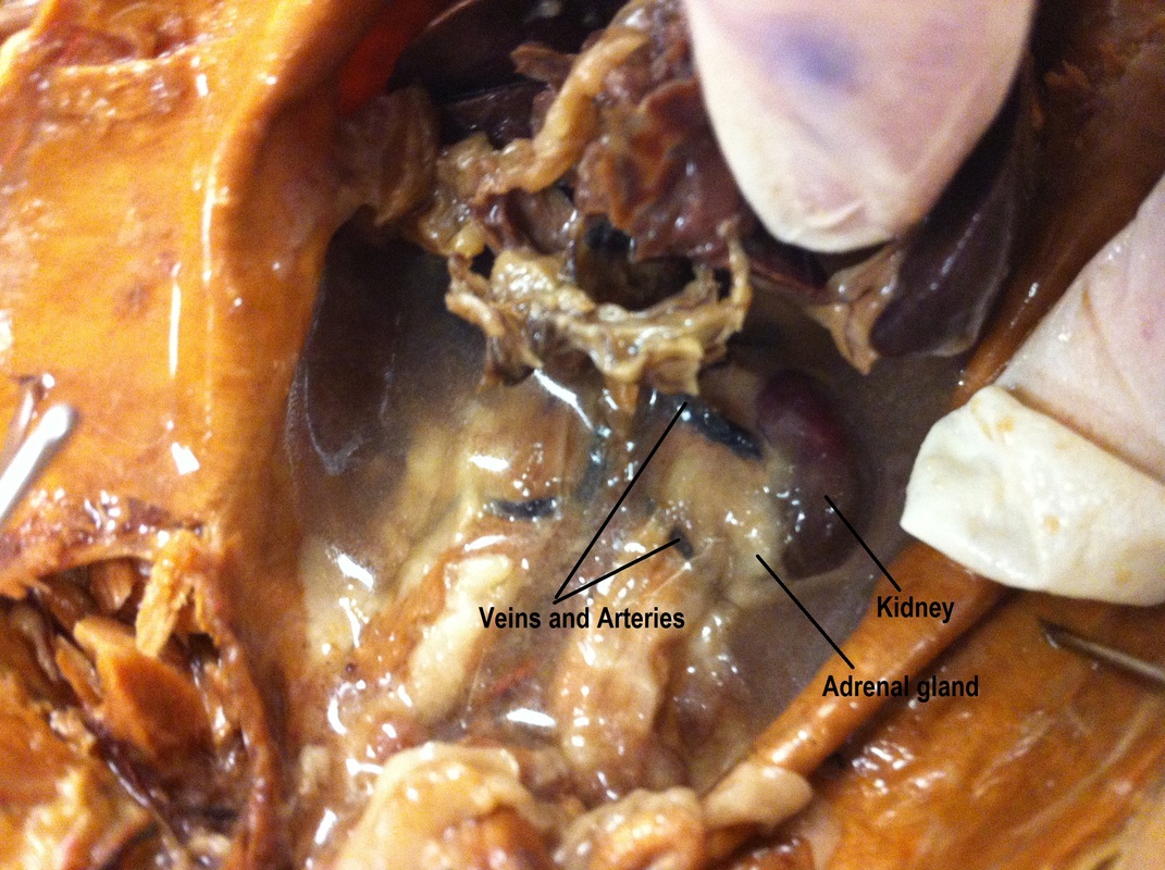
1. Locate the kidneys. Note the veins and arteries that connect with the kidneys.
2. The small yellowish glands embedded in the fat atop the kidneys are the adrenal glands which secrete adrenaline into the blood during times of crisis
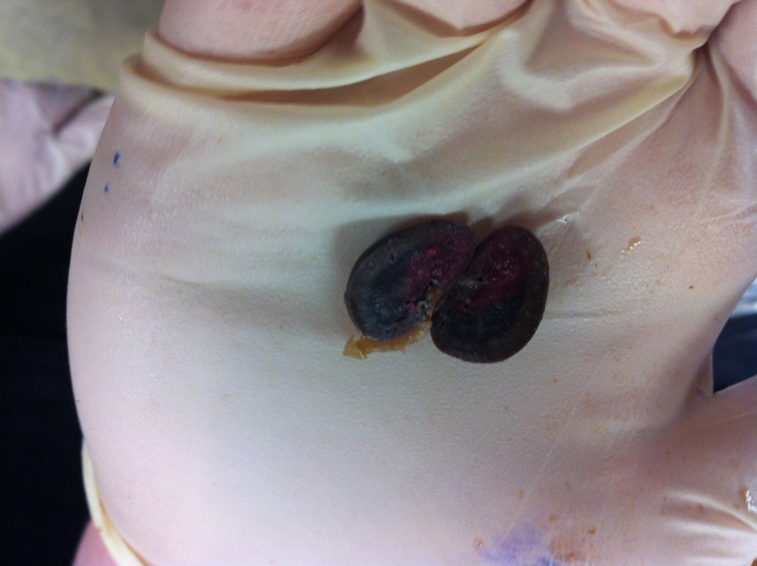
3. Remove one of the kidneys and cut it lengthwise. Notice the very fine veins and arteries within. Blood is filtered through the kidneys approximately once every 45 minutes.
Part 7: Reproductive systems
MALE (our rat, the one pictured)
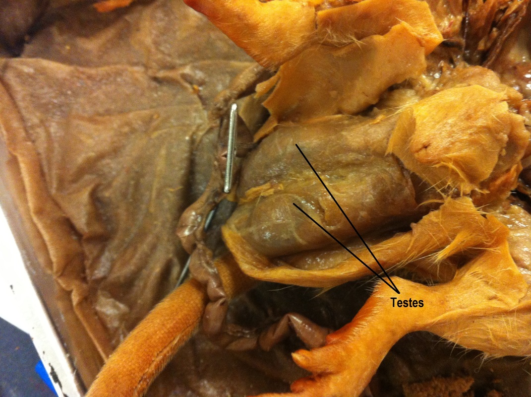
1. The major reproductive organs of the male rat are the testes, located in the scrotal sac. Cut through the sac carefully to reveal the testes. On the surface of the testes is a coiled tube called the epididymus, which collects and stores sperm cells. The tubular vas deferens moves sperm from the epididymus to the urethra, which carries sperm through the penis and out of the body.
2. The lumpy brown glands located to the left and right of the urinary bladder are the seminal vesicles. The gland below the bladder is the prostate gland, which is partially wrapped around the penis. These two things secrete materials that form the semen.
FEMALE
1. The short gray tube lying dorsal to the urinary bladder is the vagina. It divides into two uterine gorns that extend towards the kidneys. This duplex uterus is common in some animals and will accomodate multiple embryos (a litter). In contrast, a simple uterus, like in humans, has a single chamber for development of one embryo
2. At the tips of the uterine horns are small lumpy glands called ovaries, connected to the uterine horns via oviducts. Oviducts are very small, and may not be visible without a dissecting microscope.
Part 8: Circulatory system
1. The general structure of the circulatory system of the rat is almost identicle to that of humans. Pulmonary circulation carries blood through the lungs for oxygenation and then back to the heart. Systemic circulation moves blood through the body after it has left the heart.
2. Your rat specimen has been double ejected with latex to indentify veins and arteries (in our rat, the latex is not at all visible, except in the pulmonary artery). Veins carry used blood (blue) back to the heart and lungs. The lungs reoxygenate the blood and the heart pumps it back into the rest of the body. In the human body, these veins are not the same bright blue seen in the rat. However, if you look at your arm, you can see some bluish veins close to the skin. Look in your rat for veins.
3. The arteries are stained red. Find the arteries.
4. Remove the heart from the pericardial sack. You need to sever the arteries and veins connecting the heart to the circulatory system. Do not cut more than necessary. Leave as much of the veins and arteries attached to the heart as possible.
5. Identify the aorta, left and right atrium, and left and right ventricle. Carefully insert a probe into these openings and into the centre of the heart.
6. Make an incision between the left and right ventricles with the scalpel. Try to locate the bicuspid and semilunar valves.
Analysis
1. Check off each body part as you identify the variour structures throughout the rat.
2. Q: The sphincter is a circular muscle. Why is it this shape and what does it do?
A: The sphincter is a muscle that is at the beginning and end of both the small and large intestine. It is circular because it acts as an adapter from stomach to intestine, so it must be tube-shaped just like the intestine. Its purpose is to open and close to push food through the digestive system.
3. Q: Why is there a difference in the diameter and length of the small and large intestine?
A: The small intestine, being much longer than the large intestine, has a larger surface area. This is needed, as the majority of the nutrients from food are absorbed here. The large intestine does not have to have as large a surface area, as it is there to reabsorb water. Also, when food exits the stomach, it is watery, so it can travel through a thinner tube. Once the water and nutrients are filtered out, the waste becomes rather hard, so it needs more space to travel through.
4. Q: The liver is the largest organ in the body. What are its functions?
A: The liver produces bile to aid in digesting fats, as well as to store glycogen and to form wastes them into less harmful substances.
5. Q: In each of the cavities there is a membrane that covers both the wall of the cavity and the organ it contains. What is the function of these membranes?
A: The membranes house capillaries, which are used to take oxygenated blood to the organs and skin, as well as carry nutrients to where they are needed.
6. Q: What is the function of the spleen?
A: It is associated with the circulatory system, functioning in the destruction of blood cells as well as in blood storage. It also helps the immune system function.
7. Q: What is the function of the diaphragm?
A: The diaphragm is a muscle that aids in respiration. When breathing in, the diaphragm contracts, leaving more room for the lungs to expand, allowing more air to enter. When exhaling, the diaphram expands, compressing the lungs and forcing air out of them.
8. Q: What distinguishes the atria from the ventricles?
A: The atria are used to hold the blood when it enters the heart. The ventricle takes blood from the atrium and pumps it out of the heart to either the lungs or the body. The atria are also slightly smaller than the ventricles.
9. Q: Why is the wall of the left ventricle thicker than the right?
A: The left ventricle needs to pump blood throughout the entire body, so more muscle to produce more force is needed. The right ventricle only has to pump blood to the lungs, so less muscle is needed as less force is required.
10. Q: What similarities exist betweetn the male and female reproductive systems?
A: The adrenal gland is attatched to both the male and female reproductive systems. The bladder is also part of both systems, as well as the urethra. They have a similar shape, with the seminal vesicles in males and urterine horns and ovaries in females forming a Y-shape with the bladder and urethra.
11. Q: What do the kidneys do?
A: Every 45 minutes, blood is sent through the kidneys, where it is filtered of any contaminents collected from circulating the body.
